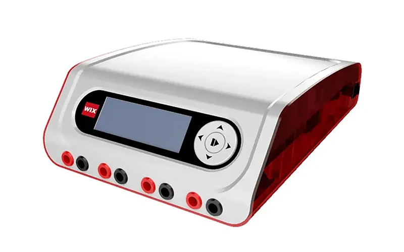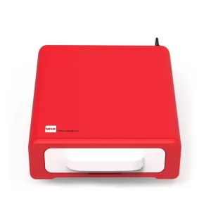
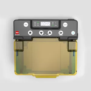
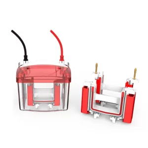
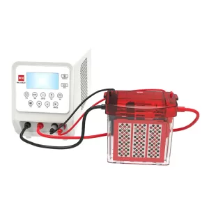
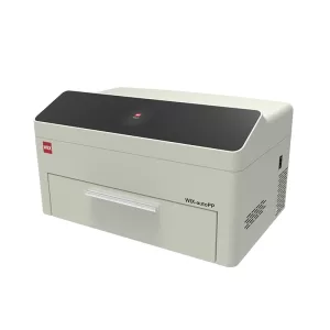
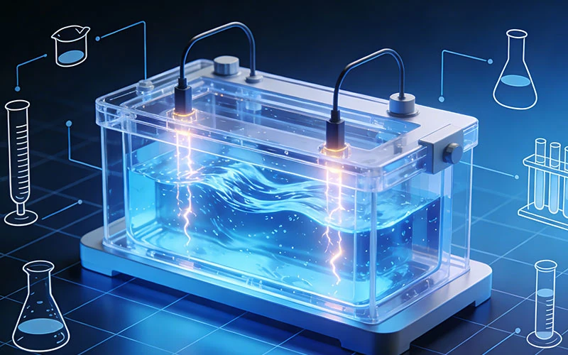
Do You Have Interest in Electrophoresis Tanks?
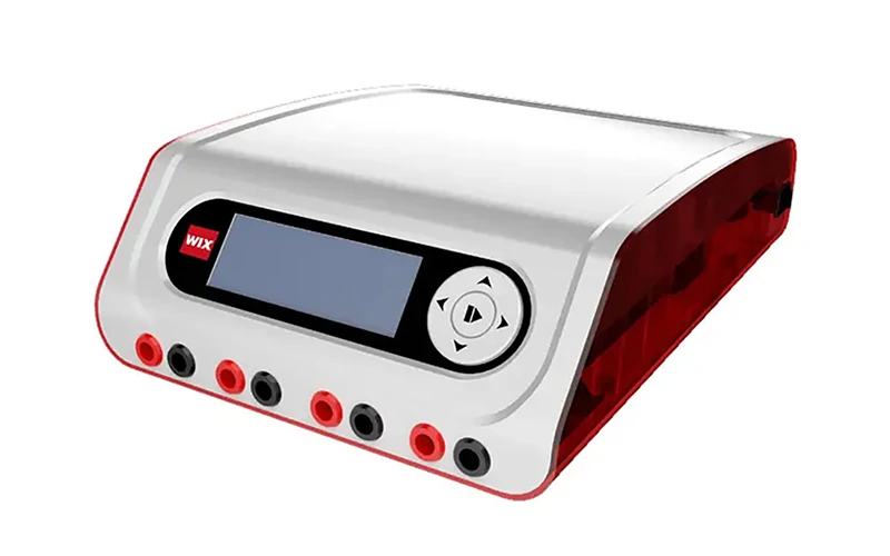
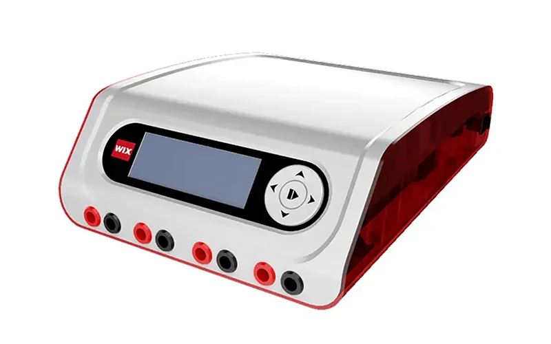
An electrophoresis tank is a core device used to hold electrophoresis buffer and samples in electrophoresis experiments. Its design and performance directly affect the accuracy and reproducibility

Key Takeaways
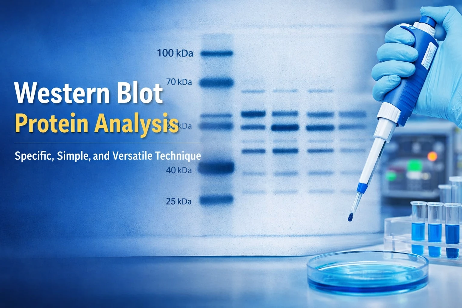
Picture a photograph taken in a completely dark room, where the subject gives off a glow so weak that your eyes cannot detect it. A Chemi Imaging System does exactly that for a Western blot. It records the faint light released when a chemical substrate reacts with a labeled antibody, then turns that light into a clean digital image.
The word “chemi” comes from chemiluminescence, which simply means light produced by a chemical reaction instead of a lamp. During a blot experiment, researchers attach an enzyme to the antibody that binds their protein of interest. Once they add the substrate, the enzyme triggers a reaction that emits photons. Those photons carry the information about where the protein sits on the membrane.
Before cameras took over, labs captured this signal on X-ray film. You would press the membrane against the film, wait for the exposure, then develop it in a darkroom. That method still works, but it costs time and material. Film offers a narrow dynamic range, each attempt uses up a sheet, and one mistake means repeating the blot. A digital setup removes most of that friction. You see the result on a screen within seconds, adjust the exposure if needed, and save the image straight to a file.
The process sounds complex, but each step makes sense once you break it into parts.
After proteins are separated on a gel and transferred onto a membrane, the membrane is incubated with antibodies. One of those antibodies carries an enzyme. When the chemiluminescent substrate arrives, the enzyme breaks it down and releases energy as light. A brighter band tells you that more of the target protein is present. The glow peaks quickly and then fades, so timing matters.
The membrane goes into a sealed, light-tight chamber. Inside, a sensitive camera looks down at it through a lens. The chamber blocks every stray photon from the room, because even a small leak of daylight would drown out the weak signal from the blot. The camera collects light over a chosen exposure, which can last from a fraction of a second to many minutes, depending on signal strength.
Software converts the collected signal into a picture you can actually read. You can view it on a monitor, adjust contrast, measure band intensity and export the result in a standard format. A good Western Blot Imaging System keeps this workflow short, so the scientist spends time on the biology rather than on the machine.
Every instrument, whatever the brand, is built from the same handful of pieces:
Heat makes electrons move inside a sensor, even in total darkness. That movement shows up as grain in the final picture. Cooling slows it down. The colder the camera runs, the longer you can expose a faint blot before the background turns noisy. This is why Gel Imaging and Documentation Systems built for research use a cooled sensor instead of an ordinary camera.
Specs can feel like alphabet soup, yet only a few numbers change what you see on the screen.
The sensor is the chip that turns light into pixels. A larger format collects more light and usually delivers a better image. Pixel count matters less than many buyers expect. A high-megapixel camera with tiny pixels can perform worse than a lower-resolution model with bigger pixels, because each small pixel captures fewer photons.
Cooling is measured as a temperature drop below the surrounding air, and a deeper drop means a quieter sensor. Bit depth describes how many shades of grey the camera can record. An 8-bit image holds 256 shades, while a 14-bit image holds more than 16,000. That extra range matters when a blot contains a very bright band next to a very faint one. You can see both without washing out the strong signal or losing the weak one.
| Spec | What it means | What to look for |
| Sensor | The chip that turns light into pixels | A larger format with reasonably large pixels |
| Resolution | Total number of pixels in the image | Enough for publication, not just the biggest number |
| Cooling | How far the sensor drops below room temperature | Deeper cooling for faint signals and long exposures |
| Bit depth | Number of grey shades the camera records | 14-bit or higher for a wide dynamic range |
| Lens aperture | How much light the lens gathers | A low f-number for faster imaging |
| Exposure range | Shortest to longest capture time | A wide range for both strong and weak blots |
An Imaging System for Western Blot should balance these numbers. One impressive figure rarely tells the whole story.
Chemi imaging started with Western blots, but the same hardware handles several jobs.
Researchers use it to detect proteins, confirm changes in gene expression and document gels. Because the output is digital, images can be measured, annotated and dropped straight into a paper or a report. That convenience is a major reason Chemiluminescence Documentation Systems have become standard equipment in life science labs.
The user base is broad. Academic and research institutes run everyday protein work. Pharmaceutical and biotech teams apply imaging to drug discovery and quality checks. Hospitals and diagnostic labs use it for a range of assays. Food safety, forensic and anti-doping labs turn to it when they need sensitive, repeatable detection.
Both methods produce light, but the source differs. Chemiluminescence comes from a chemical reaction and needs no external lamp. Fluorescence requires a light source to excite a dye, which then glows. Many modern instruments support both, giving a lab more flexibility from a single device.
The WIX-chemiPHOTO brings these pieces together in one compact unit. It is built for labs that want sensitive blot imaging without a complicated setup, and it covers the specs that matter most.
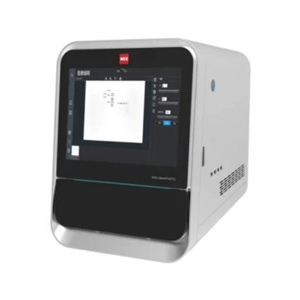
At the center sits a SONY IMX492 sensor with a 4/3-inch format and 4.63 × 4.63 µm pixels. Effective resolution reaches 4164 × 2796, which works out to an 11.7-megapixel image. Peak quantum efficiency is 90%, so the chip converts a high share of incoming photons into signal. A 14-bit A/D converter handles a full well capacity of 65 ke⁻, while readout noise measures 1.6–1.2 e⁻ at high gain and 6.9–5.2 e⁻ at low gain.
Dark current sits at just 0.002 e⁻ per pixel per second at −20 °C. Two stages of thermoelectric cooling bring the sensor to roughly 35 °C below ambient, which keeps long exposures clean. The maximum image area is 10 × 14 cm, large enough for standard mini and midi blots.
The F0.95 lens gathers plenty of light, and the unit handles auto focus and aperture adjustment on its own. Exposure times run from 60 microseconds to 3600 seconds, so the same camera covers a bright band and a barely visible one. A double-layer front door blocks stray light, and you can open and close the door with a gesture without touching it, which helps avoid cross contamination. The 12.1-inch capacitive touch screen and built-in computer put the controls in one place.
Image analysis software comes with the unit at no extra cost. It handles protein blot imaging and helps prepare figures for publication. The system holds a CE certificate (IT1440WX08111806) and an ISO 9001 certificate (53123QZ0615R0S). You will find it in academic research, pharmaceutical and biotech labs, clinical diagnostics, food safety and forensic work.
It captures light from chemiluminescent reactions, most often on a Western blot membrane. Labs use it to detect proteins, document gels and produce images for reports or publication.
An enzyme attached to an antibody reacts with a substrate and releases faint light. A cooled camera inside a dark chamber records that light, and software turns the signal into a digital image.
Chemiluminescence produces light through a chemical reaction and needs no lamp. Fluorescence relies on an external light source to excite a dye. Many systems can handle both methods.
It varies with signal strength. A strong band may need only a second or two, while a faint one can take several minutes. The exact window depends on the substrate and the camera.
Focus on the sensor, cooling depth, bit depth and lens aperture. Check that the software is easy to use and that the camera can handle both strong and weak signals.
A chemi imaging system takes the guesswork out of blot detection. Instead of darkrooms and film, you get a repeatable digital workflow that shows faint signals clearly and saves every result. Understanding a handful of specs, namely the sensor, cooling, bit depth and lens, is enough to tell a capable instrument from a weak one. The WIX-chemiPHOTO puts those specs together in a single, straightforward unit for labs that want dependable imaging day after day.
Request a quote or a detailed data sheet to see whether the WIX-chemiPHOTO fits your lab.