Western blot, also known as immunoblotting, is a widely used laboratory technique for the detection and analysis of specific proteins in a complex biological sample. It combines gel electrophoresis for separating proteins by molecular weight with immunodetection using specific antibodies to identify target proteins. Here is a detailed overview of the key steps, principles, and applications of Western blotting:
1. Sample Preparation
The first step involves extracting proteins from the biological sample (e.g., cells, tissues, or fluids). This typically requires homogenization in a lysis buffer containing detergents (e.g., SDS) to solubilize proteins, protease inhibitors to prevent protein degradation, and phosphatase inhibitors (if studying phosphorylated proteins). The lysate is then centrifuged to remove cellular debris, and the protein concentration is quantified (e.g., using BCA or Bradford assay) to ensure equal loading in subsequent steps.
Tissue grinders are essential for extracting nucleic acids (DNA, RNA), proteins, and metabolites from biological samples. Bead mill grinders, in particular, are widely used in genomics and proteomics to disrupt tissues into uniform lysates, enabling downstream applications like PCR, Western blotting, and next-generation sequencing. Cryogenic grinding ensures RNA integrity by preventing RNase activity during tissue disruption.
WIX-3dGRIND 3-Dimension Tissue Grinder and WIX-manyGRIND Tissue Grinder will meet your demands, with 12x2ml/24x(0.2-0.5ml) /24x2ml samples within 3 minutes.
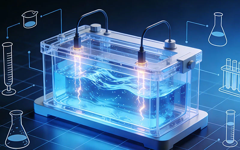
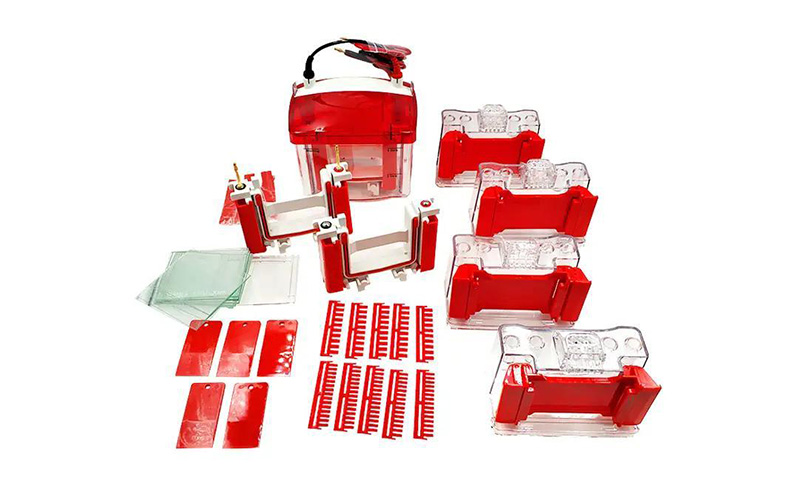
2. Gel Electrophoresis (SDS-PAGE)
Proteins are separated by size using Sodium Dodecyl Sulfate-Polyacrylamide Gel Electrophoresis (SDS-PAGE). SDS, an anionic detergent, denatures proteins and coats them with a uniform negative charge, ensuring separation is based primarily on molecular weight (smaller proteins migrate faster through the polyacrylamide gel matrix). A stacking gel (low acrylamide concentration) first concentrates proteins into a tight band, followed by a resolving gel (higher acrylamide concentration) for size-based separation. After electrophoresis, proteins appear as discrete bands in the gel, with molecular weight markers (ladder) run alongside to estimate target protein size.
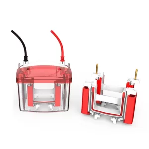
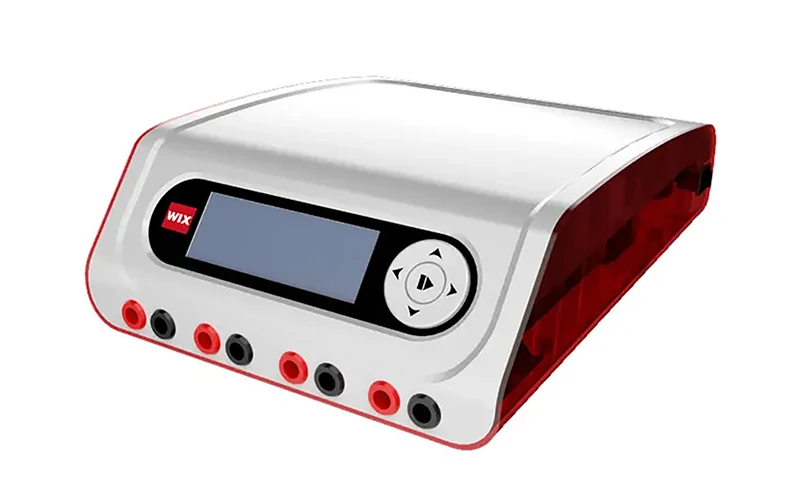
WIX-miniPRO series and WIX-easyPRO series includes 2-gel and 4-gel electrophoresis. Details you can refer to product page.
3. Protein Transfer (Blotting)
Proteins are transferred from the gel to a solid support membrane (typically nitrocellulose or PVDF) via electrophoresis (electroblotting). This step is critical for subsequent antibody binding, as membranes provide a stable, accessible surface for immunodetection. Transfer conditions (e.g., voltage, time, buffer) are optimized to ensure efficient and uniform transfer of proteins from the gel to the membrane. After transfer, the membrane is briefly stained (e.g., with Ponceau S) to confirm equal loading and successful transfer of protein bands.
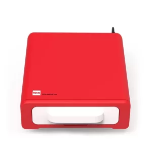
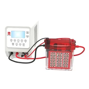
WIX TECHNOLOGY offers wet transfer of 2-gel and 4-gel and semi-dry transfer that meet various of demand. Moreover, WIX TECHNOLOGY also has other choices, such as Fast Wet Blotter WIX-coolBLOT, with built-in power supply and buffer cooling that there is no need to connect an extra power supply and cooling the buffer by the ice. For more details, you can refer to its product page.
4. Blocking
To prevent non-specific binding of antibodies to the membrane, the membrane is incubated in a blocking buffer containing proteins (e.g., bovine serum albumin, BSA, or non-fat milk) and detergents (e.g., Tween-20). Blocking typically occurs for 1–2 hours at room temperature or overnight at 4°C, ensuring that unoccupied sites on the membrane are saturated with non-specific proteins.
For manual blocking, shakers are recommended.
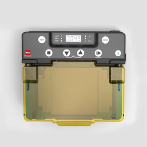
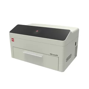
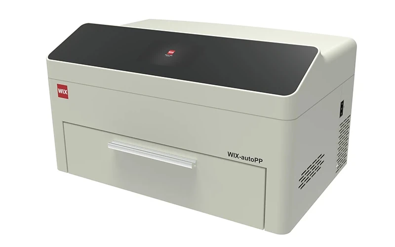
Automatic Western Blot Processing System WIX-autoPP can finish incubation and blocking of antibodies automatically.
5. Primary Antibody Incubation
The membrane is incubated with a primary antibody that specifically recognizes the target protein. Primary antibodies are either monoclonal (recognizing a single epitope) or polyclonal (recognizing multiple epitopes) and are diluted in blocking buffer to minimize non-specific binding. Incubation conditions (e.g., concentration, time, temperature) vary depending on antibody affinity and are optimized to maximize specific binding. After incubation, the membrane is washed extensively with a wash buffer (e.g., TBST) to remove unbound primary antibody.
Automatic Western Blot Processing System WIX-autoPP can finish incubation and blocking of antibodies automatically.
6. Secondary Antibody Incubation
The membrane is then incubated with a secondary antibody that binds to the primary antibody. Secondary antibodies are conjugated to a detectable label, such as horseradish peroxidase (HRP) or alkaline phosphatase (AP), or fluorescent dyes. This step amplifies the signal, as multiple secondary antibodies can bind to a single primary antibody. After incubation, the membrane is washed again to remove unbound secondary antibody.
Automatic Western Blot Processing System WIX-autoPP can finish incubation and blocking of antibodies automatically.
7. Detection
The labeled secondary antibody is visualized using a detection method compatible with its label:
- Chemiluminescence: HRP-conjugated secondary antibodies react with substrates (e.g., ECL) to produce light, which is detected using X-ray film or a digital imager.
The resulting bands correspond to the target protein, with band intensity proportional to protein abundance (semi-quantitative analysis).
WIX-chemiPHOTO Chemi imaging system, an integrated fully automatic chemiluminescence imaging system,based on technology of advanced scientific grade cameras and lenses, accommodating both automatic and manual imaging modes, and a built-in computer for fast imaging.
WIX-multiPHOTO Multi-functions Gel Doc, an integrated fully automatic imaging system of chemiluminescence and Ultraviolet, based on technology of advanced scientific grade cameras and lenses, and a built-in computer for fast imaging.
8. Quantification and Analysis
Band intensity is quantified using image analysis software (e.g., ImageJ, Quantity One). To control for loading variability, target protein levels are normalized to a housekeeping protein (e.g., β-actin, GAPDH) that is constitutively expressed and not affected by experimental conditions. This allows for semi-quantitative comparison of protein expression across samples.
Applications
Western blotting is widely used in molecular biology, biochemistry, and clinical research for:
- Detecting the presence or absence of a specific protein in a sample.
- Semi-quantifying protein expression levels.
- Characterizing protein modifications (e.g., phosphorylation, glycosylation) via epitope-specific antibodies.
- Validating antibody specificity or protein expression in knockout/knockdown models.
- Diagnosing diseases (e.g., HIV, Lyme disease) by detecting pathogen-specific proteins or autoantibodies.
Limitations
- Semi-quantitative: Results are relative and require normalization to housekeeping proteins; absolute quantification is not possible.
- Sensitivity: Less sensitive than ELISA or mass spectrometry, requiring relatively high protein concentrations.
- Reproducibility: Prone to variability from factors like antibody quality, transfer efficiency, and blocking conditions.
Despite these limitations, Western blotting remains a cornerstone technique for protein analysis due to its specificity, simplicity, and versatility.