Do You Have Interest in Electrophoresis Tanks?
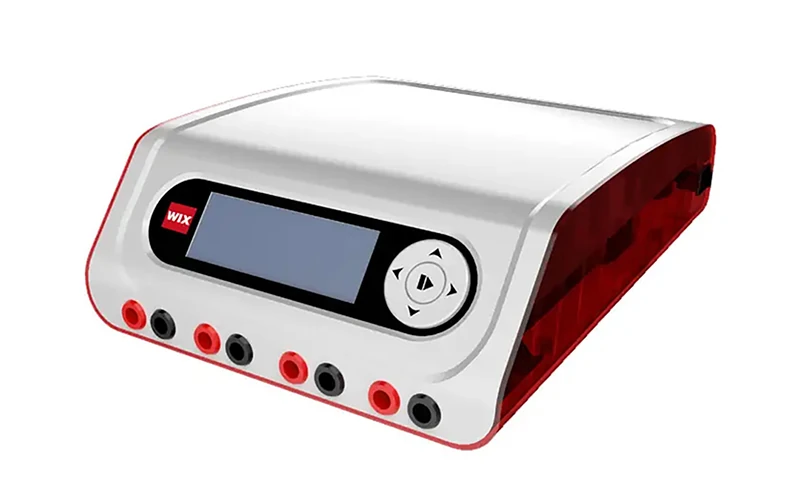
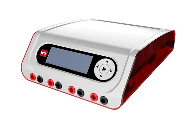
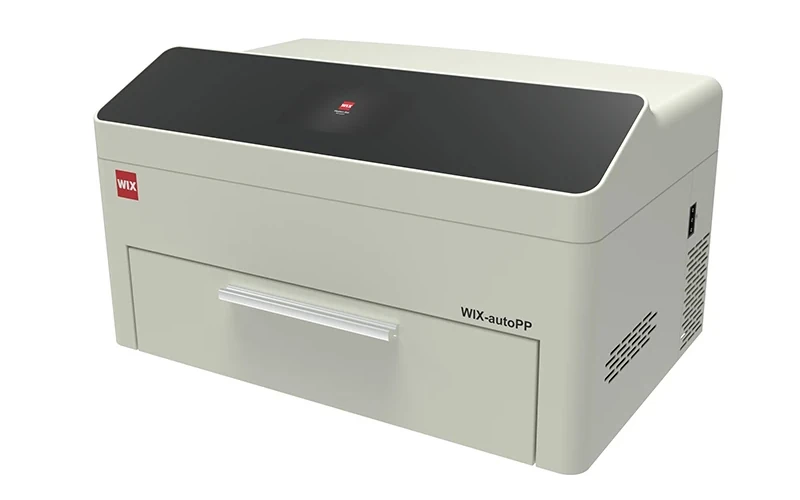
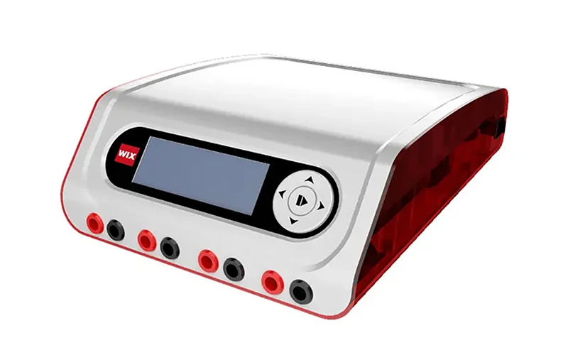
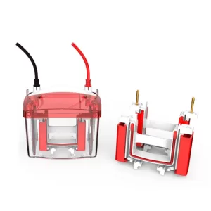
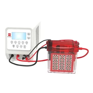
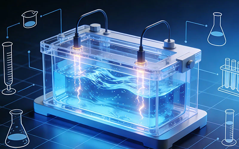
An electrophoresis tank is a core device used to hold electrophoresis buffer and samples in electrophoresis experiments. Its design and performance directly affect the accuracy and reproducibility

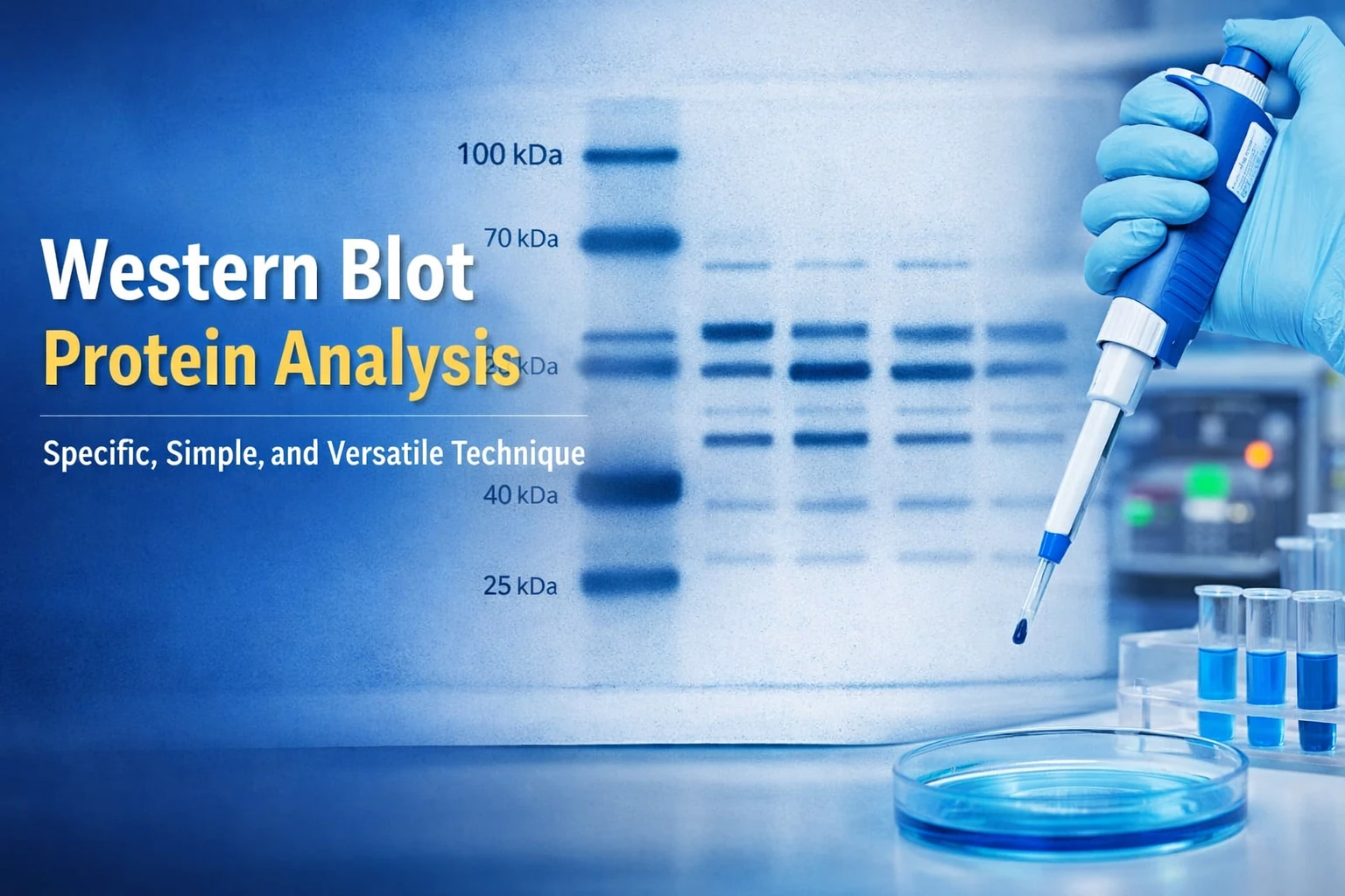
Electrophoresis gels and Western blots generate visual evidence that supports molecular biology, biotechnology, and biomedical research. However, observing bands during an experiment is only one part of the workflow. Laboratories also need to preserve experimental records, compare results, interpret band patterns, and support reproducibility across experiments.
A Gel Documentation system provides an integrated imaging workstation for capturing and recording these results digitally. By combining controlled illumination, optical components, a sensitive camera, and image-processing software, a gel documentation system converts visible or chemically generated sample signals into digital images suitable for documentation and, where supported, quantitative analysis.
The imaging process generally follows a sequence: sample illumination or light generation, optical filtering, photon collection, camera capture, and software-based image processing. Different detection methods, such as fluorescence and chemiluminescence, require different optical and sensitivity configurations.
This guide explains how gel documentation systems work, compares imaging technologies, reviews important technical specifications, and outlines purchasing considerations. It also introduces WIX-multiPHOTO as an integrated imaging platform for laboratories evaluating flexible gel and Western blot imaging workflows.
A gel documentation system is an enclosed digital imaging workstation designed to capture images of electrophoresis gels and other compatible biological samples. It typically integrates an imaging chamber, illumination sources, optical filters, a camera, and software into a controlled operating environment.
In conventional laboratory workflows, researchers may use a transilluminator for visual inspection and a separate camera for recording images. A dedicated gel imaging system combines these functions into a more consistent workflow, reducing dependence on room lighting, manual camera positioning, and improvised image capture arrangements.
Controlled imaging conditions can improve repeatability by standardizing sample positioning, illumination, exposure settings, and image storage. These controls are particularly useful when laboratories need to compare results across multiple experiments or maintain traceable experimental records.
The system does not eliminate the need for appropriate sample preparation or experimental controls. Instead, it provides a more structured way to capture the signals produced by those experiments.
Although the terms are often used interchangeably, they describe different stages of the workflow.
| Term | Meaning | Main purpose |
| Gel documentation | Recording and preserving experimental images | Creating a digital experimental record |
| Gel imaging | Optically capturing signals from a sample | Converting fluorescence, reflected light, or chemiluminescence into an image |
| Gel analysis | Measuring and interpreting image features | Evaluating band intensity, relative signal, or other measurable characteristics |
A basic transilluminator may illuminate a gel without providing a complete digital capture or analysis workflow. Similarly, a camera-equipped gel imaging system may document images without offering validated quantitative measurements for every application.
Therefore, the presence of imaging software does not automatically establish that a system provides accurate quantitative analysis.
Gel documentation equipment is commonly used to record and review results from a range of molecular biology and biochemical workflows. Typical applications include:
The required imaging configuration varies according to the sample type, detection chemistry, signal strength, and whether the workflow requires simple documentation or quantitative analysis. Laboratories should therefore evaluate application compatibility before selecting a system rather than relying on camera specifications alone.
A gel documentation system converts sample-derived optical signals into a digital image through several coordinated stages.
The operator places the stained gel, membrane, or other compatible sample on the imaging stage. Proper positioning ensures that the sample remains within the usable imaging area and that the camera can capture the relevant region without obstruction.
Sample preparation and positioning affect image quality. Excessive background staining, uneven sample thickness, bubbles, dust, or unsuitable containers may introduce artifacts or obscure bands. The imaging stage should accommodate the dimensions and handling requirements of the intended samples.
The operator selects an illumination mode according to the detection chemistry. Common modes include UV illumination, blue-light excitation, white-light illumination, and epi-illumination.
The selected illumination source should match the optical requirements of the sample and the detection method. For fluorescence imaging, the excitation wavelength should be compatible with the stain or fluorophore being used. For transmitted or reflected-light imaging, the illumination geometry should provide sufficient contrast for the sample.
The specific illumination configurations and their application considerations are discussed in the Imaging Technologies section below.
A wavelength that is unsuitable for the sample may produce weak fluorescence, poor contrast, or excessive background.
An inappropriate wavelength may lead to a weakened or even absent fluorescence signal, and may also result in a decrease in contrast or a higher background level.
UV illumination also requires appropriate safety controls, including an enclosed imaging chamber, interlocks where applicable, and operating procedures that limit unnecessary UV exposure.
The optical signal reaching the camera depends on the imaging mechanism.
Fluorescence imaging: A fluorophore absorbs excitation light and subsequently emits light at longer wavelengths. The emitted signal is collected by the optical system and separated from unwanted excitation light using suitable filters.
Reflected-light imaging: The camera detects light reflected from the sample surface. The contrast depends on the sample’s optical properties, staining characteristics, illumination geometry, and background.
Chemiluminescence imaging: A chemical reaction generates light without requiring external excitation in the same manner as fluorescence. Chemiluminescent Western blot imaging therefore depends on detecting relatively weak light produced by the reaction.
These mechanisms are not interchangeable. A system designed for fluorescent gel imaging may require additional optical and camera capabilities to support chemiluminescence effectively.
Optical filters help separate useful emission signals from unwanted excitation light and other background components. In fluorescence imaging, an emission filter is selected to transmit the wavelengths emitted by the fluorophore while reducing the passage of excitation light.
The filter’s transmission range, optical quality, and compatibility with the illumination source influence the quality of the captured signal. Standard and optional filters should be evaluated against the dyes and stains used in the laboratory.
Lens design is another important factor. A large-aperture lens can collect more light, which is particularly useful when imaging weak fluorescence or chemiluminescent signals. However, aperture alone does not determine overall system performance. Sensor sensitivity, optical transmission, focus, and background control also contribute.
High-quality AR+AR optical windows, combined with multi-layer anti-reflection coatings, can reduce unwanted reflections and optical losses. Such design features may improve signal transmission and image clarity when appropriately integrated into the imaging path.
Photons collected by the optical system reach the camera sensor, where they are converted into electronic signals and then digital pixel values. The resulting image represents the spatial distribution and intensity of the detected light.
Exposure duration influences the amount of signal accumulated by the sensor. Longer exposures may help capture weak signals, while shorter exposures can reduce the risk of saturation when signals are strong.
Electronic rolling-shutter operation is one possible camera readout method. With a rolling shutter, different rows of the sensor are read sequentially rather than all at exactly the same instant. For stationary gel and blot imaging, this may be suitable, but the camera’s readout characteristics should still be considered alongside exposure control and application requirements.
Saturation occurs when a pixel reaches the upper limit of its measurable signal range. Once saturated, additional signal cannot be represented accurately in that pixel. Maintaining useful signal levels within the sensor’s measurable range is essential for preserving image information and supporting reliable intensity comparisons.
After capture, software displays and processes the image. Common functions include contrast adjustment, background correction, image cropping, band detection, densitometry, and digital archiving.
Display enhancement should be distinguished from modification of the underlying quantitative data. Adjusting brightness or contrast may improve visual interpretation, but it does not necessarily improve measurement accuracy. Quantitative workflows should retain the original image and document the processing steps used for analysis.
Depending on the software, users may export images in formats such as TIFF, PNG, or other supported formats. For quantitative applications, preserving original image data and acquisition metadata is important. Export settings should not unintentionally reduce bit depth or introduce processing that compromises subsequent analysis.
Gel imaging systems may use different illumination configurations depending on the optical properties of the sample and the detection chemistry. The main difference between these modes is how light is delivered to the sample and how the resulting signal is collected.
Typical configuration
| Illumination | Representative configuration | Primary consideration |
| UV | 365 nm or 302 nm | Match the excitation wavelength to compatible fluorescent stains while maintaining appropriate UV safety controls |
| Blue-light | 470 nm | Match the excitation spectrum of the fluorescent label and use an appropriate emission filter |
| White-light | Transmitted or reflected white light | Suitable for samples where contrast is generated through staining or differences in optical properties |
| Epi-illumination | Illumination through an optical path above or around the sample | Useful when the sample requires illumination from the imaging side rather than transmission through the sample |
UV illumination can provide excitation for compatible fluorescent stains, but the selected wavelength should correspond to the excitation characteristics of the stain. Enclosed imaging chambers and appropriate safety mechanisms are important when UV sources are used.
Blue-light illumination is an alternative for compatible fluorescent labels. Because the excitation wavelength differs from UV, the corresponding emission filter must also be suitable for the fluorophore being detected.
White-light imaging relies on transmitted or reflected light rather than fluorescence excitation. It can therefore be useful for stained samples whose visual contrast does not depend on fluorescence.
Epi-illumination uses a different optical geometry in which light is directed toward the sample through the imaging optical path. Its suitability depends on sample structure, signal generation, and the configuration of the imaging system.
The stain or fluorophore determines which excitation and emission wavelengths are appropriate. A gel documentation system should therefore be evaluated as a complete optical configuration rather than by camera specifications alone.
Important considerations include:
Begin by identifying your lab’s current and future sample types, stains, fluorophores, and detection methods.
Always request a detailed, itemized configuration list distinguishing standard hardware (chamber, camera, computer, default illumination, and standard emission filters) from optional add-ons (extra filters, specialized sample trays). This prevents unexpected post-purchase costs and ensures immediate operational readiness.
Equipment pricing is driven by camera technology, sensor performance, illumination modes, and software integration. When budgeting, evaluate the total cost of ownership, which includes:
Verify that prospective suppliers provide transparent technical specifications, user manuals, and compliance documentation. While quality certificates support vendor evaluation, they do not replace application-specific validation.
Before ordering, confirm site requirements (power, environment, network connectivity). Define clear acceptance criteria—such as image uniformity, sensitivity, and software functionality—during installation. Finally, document warranty terms, service response times, and training commitments to ensure long-term operational continuity.
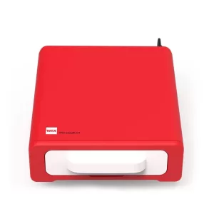
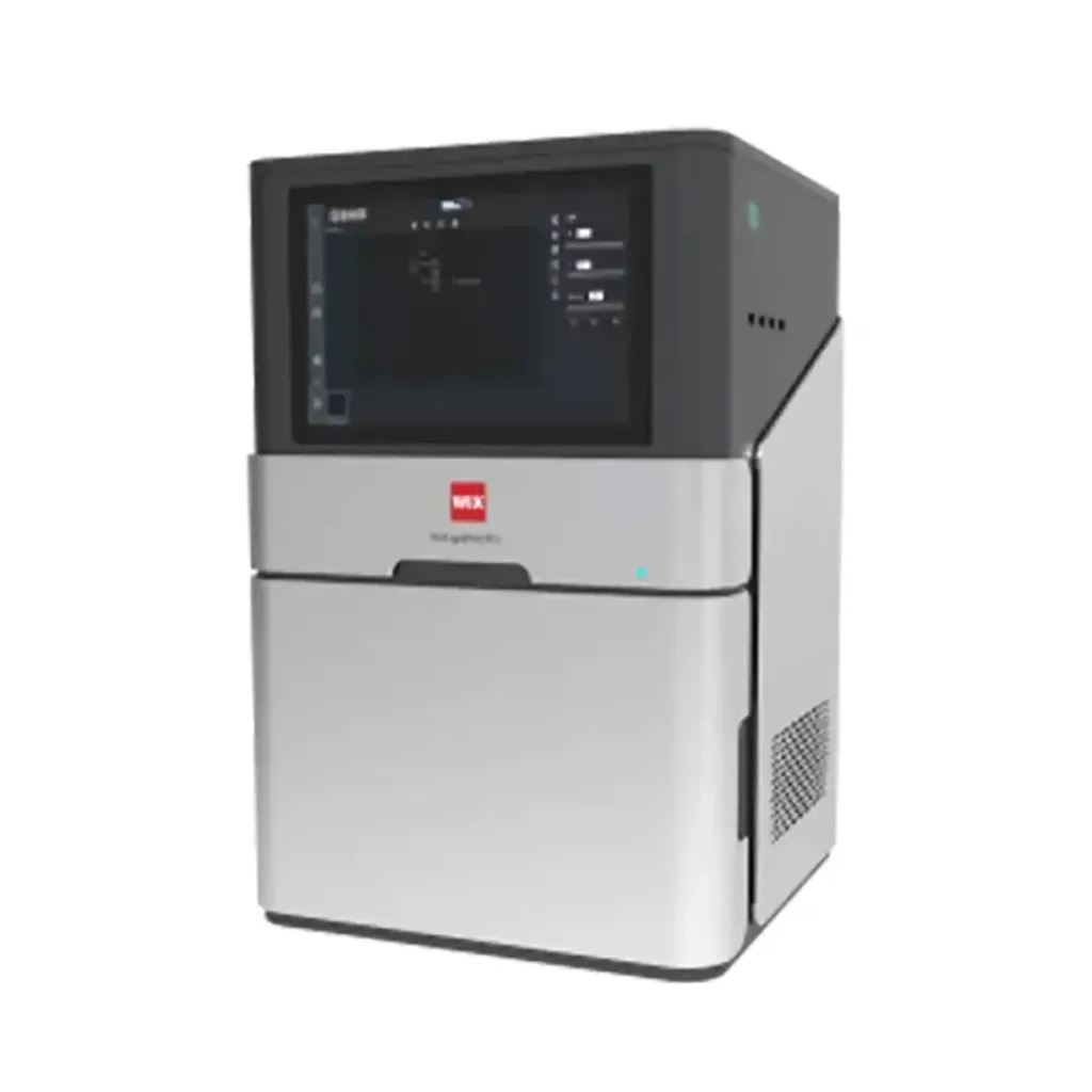
WIX-multiPHOTO is presented as an all-in-one flexible imaging platform designed to support chemiluminescence and UV gel imaging within one workstation.
Its double-layer front-door light-avoidance structure is intended to reduce background stray light entering the imaging chamber. Controlling unwanted ambient light is particularly relevant when capturing weak signals, although overall image quality also depends on the camera, optics, exposure settings, and sample preparation.
An integrated design can simplify laboratory workflows by combining imaging hardware and control functions into one system. Its suitability should be evaluated against the applications and performance requirements of the intended users.
The WIX-multiPHOTO configuration includes trans-white, trans-UV, and epi-blue imaging modes.
Representative illumination specifications include:
These modes provide flexibility for different sample and detection chemistries. The selected mode must still be matched to the excitation requirements of the stain or fluorophore and the optical characteristics of the sample.
The platform includes two standard emission filters and four optional filters. This configurable filter arrangement may help laboratories adapt the system to different fluorescence applications.
WIX-multiPHOTO incorporates a built-in computer and a 12.1-inch capacitive touch LCD screen. An integrated workstation can simplify operation by reducing the need for separate control hardware and providing a unified interface for image acquisition and software functions.
The practical value of an integrated interface depends on software usability, supported analysis tools, image storage, export functions, and operator training. Laboratories should evaluate these functions through a product demonstration or technical documentation when possible.
The platform includes a SONY IMX492 sensor, TEC cooling, a large-aperture lens, and flexible illumination options.
A cooled sensor may help manage thermal noise during suitable low-light imaging workflows, while a large-aperture lens can support light collection. The usefulness of these features depends on the sample signal, required sensitivity, exposure duration, imaging area, and analysis requirements.
For laboratories evaluating WIX-multiPHOTO or any other advanced gel electrophoresis visualization machine, the relevant question is whether its integrated camera, optical configuration, illumination modes, and software support the specific assays that need to be documented. Application-specific suitability should be confirmed through technical specifications, and where appropriate, representative imaging tests.
A gel documentation system records and evaluates images of DNA, RNA, and protein gels, fluorescent samples, colorimetric results, and chemiluminescent Western blots where supported. It provides digital records for experimental interpretation, comparison, and archiving.
Fluorescence uses external excitation light, while chemiluminescence generates light through a chemical reaction. The system collects the signal through optics and filters, captures it with a camera, and processes the image using software. Exposure control is important for both signal visibility and avoiding saturation.
Compare sensor sensitivity, dynamic range, bit depth, lens aperture, filters, imaging area, exposure control, cooling, software, safety features, and upgradeability. Confirm that the complete configuration supports the intended stains and detection methods.
Cost varies with camera technology, illumination modes, chemiluminescence capability, filters, software, computer integration, accessories, and service arrangements. Obtain verified supplier quotations that clearly identify included and optional components.
A gel documentation system integrates illumination, optics, camera capture, and software to preserve visual evidence from electrophoresis gels, Western blots, and other compatible samples. Understanding the imaging chain helps laboratories distinguish basic image recording from quantitative analysis and identify the technical features that influence performance.
When selecting gel documentation equipment, consider sample type, detection chemistry, sensor sensitivity, dynamic range, imaging area, filter compatibility, software capabilities, and safety requirements. Configuration flexibility, supplier documentation, installation support, and after-sales service are also important for long-term laboratory use.
For laboratories evaluating an integrated platform such as WIX-multiPHOTO, reviewing the available imaging modes, optical configuration, camera specifications, can help establish whether the system aligns with current and future workflows. Contact the supplier for configuration details and application-specific inquiries.