Do You Have Interest in Electrophoresis Tanks?
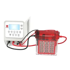
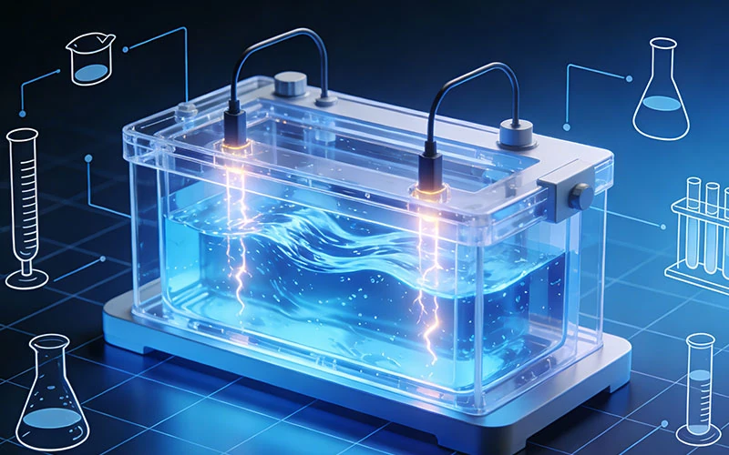
An electrophoresis tank is a core device used to hold electrophoresis buffer and samples in electrophoresis experiments. Its design and performance directly affect the accuracy and reproducibility

DNA gel electrophoresis is a routine method for separating DNA fragments according to size. DNA samples are loaded into an agarose gel, moved through the gel matrix by an electric field, and then visualized with a suitable imaging method.
A horizontal electrophoresis system allows agarose gels to be cast and run in a compact tray while supporting multiple samples. When compatible fluorescent stains are combined with blue-light illumination, DNA bands can be visualized without relying on direct UV illumination.
For laboratories selecting or upgrading electrophoresis equipment, performance depends on more than the tank itself. Agarose concentration, buffer preparation, voltage, sample capacity, heat control, staining chemistry, and imaging compatibility all affect the workflow. This guide covers gel preparation, electrophoresis, blue-light visualization, troubleshooting, maintenance, and system selection, using WIX-blueDNA as an example.
· A horizontal electrophoresis system supports agarose gel casting and DNA separation.
· Agarose concentration, buffer, voltage gradient, and electrode spacing affect migration and band resolution.
· Blue-light-compatible fluorescent stains can provide an alternative to UV-based visualization.
· WIX-blueDNA combines horizontal electrophoresis, a built-in power supply, blue-light imaging, and gel cutting.
· Gel, buffer, electrical connections, stain, and imaging compatibility should be checked before each run.
Direct UV exposure can affect DNA, especially when a band needs to be recovered for further testing. Blue light provides an alternative when the fluorescent stain is compatible with blue-light excitation.
Performance depends on the stain, illumination wavelength, optical filter, and imaging setup. These components should therefore be matched before adopting a workflow.
The electrophoresis tank provides the electric field for DNA separation, while the blue-light unit provides excitation for compatible fluorescent stains. Combining these functions can reduce gel transfers between devices.
A blue-light workflow can reduce reliance on UV transillumination and simplify gel handling when visualization and cutting are available on the same platform.
Blue light does not replace laboratory safety procedures. Operators should follow equipment instructions, use appropriate PPE, handle stains according to their SDS, and dispose of gels and buffers according to institutional requirements.
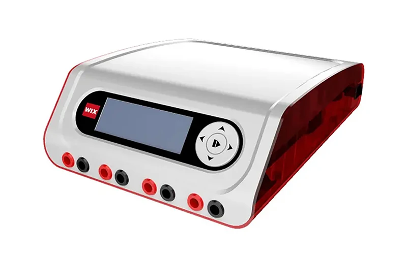
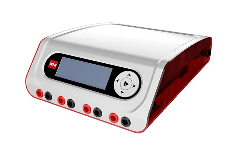
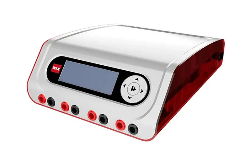
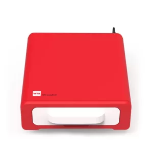
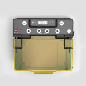
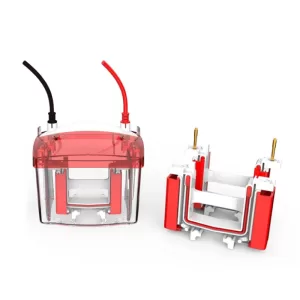
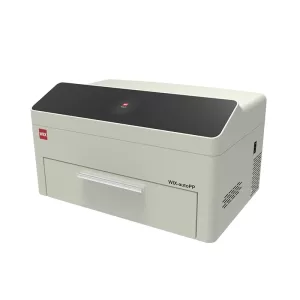
The WIX-blueDNA combines horizontal electrophoresis, a built-in power supply, blue-light imaging, and blue-light gel cutting.
When selecting a system, consider gel area, buffer capacity, voltage and current range, electrode construction, comb configuration, and power controls. WIX-blueDNA provides four gel-size configurations and up to 1,000 mL buffer capacity.
| Specification | WIX-blueDNA |
| Voltage range | 10–200 V |
| Current range | 10–500 mA |
| Maximum power | 100 W |
| Gel sizes | 120 × 120, 120 × 60, 60 × 120, 60 × 60 mm |
| Buffer capacity | Up to 1,000 mL |
| Electrode | Ø0.25 mm platinum |
| Dimensions | 300 × 170 × 80 mm |
| Weight | 2.0 kg |
These specifications allow the system to support different gel sizes and sample configurations.
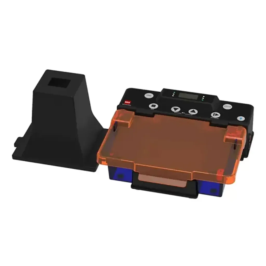
Blue-light visualization depends on matching the illuminator with the fluorescent stain and optical filter. An appropriate amber or orange filter may improve contrast with compatible stains.
WIX-blueDNA provides three blue-light intensity levels, allowing illumination to be adjusted according to stain response and image contrast.
Common blue-light-compatible stains include SYBR Safe and GelGreen, but compatibility depends on the stain and imaging system. Ethidium bromide should not automatically be assumed to provide the same response under blue light as under UV.
1X TAE and 1X TBE are commonly used electrophoresis buffers. Accurate dilution is important because buffer concentration affects conductivity, DNA migration, and heat generation.
Agarose concentration affects the pore size of the gel. Higher concentrations generally provide better separation of smaller fragments, while lower concentrations are more suitable for larger fragments.
| Approximate DNA Size | Typical Agarose Range |
| ~100–500 bp | 1.5–2.0% |
| ~500–2,000 bp | 1.0–1.5% |
| ~2,000–10,000 bp | 0.7–1.0% |
These ranges are starting points rather than fixed requirements. Final selection depends on fragment size and the resolution required.
For routine agarose gel electrophoresis, the gel concentration should match the expected DNA fragment range.
Prepare 1X TAE, 1X TBE, or another selected buffer at the required working concentration. Incorrect dilution can change migration behavior and increase heating.
Use consistent buffer conditions when comparing repeated runs.
Secure the gel tray, position the comb correctly, pour the agarose, and allow it to solidify before removing the comb. Inspect the wells for damage before loading samples.
The WIX-blueDNA uses a gel tray specified to withstand temperatures up to 100°C without deformation. This is a product-specific feature rather than a general requirement for all electrophoresis trays.
Prepare samples with the appropriate loading buffer and include a molecular-weight ladder when fragment-size estimation is required. Lower the pipette tip carefully into each well and avoid excessive sample volume.
WIX-blueDNA can use a 1 mm comb with 25 wells per row and four rows, allowing up to 100 samples including markers according to the supplied specifications.
Voltage should be considered in relation to electrode distance:
Voltage gradient = Applied voltage ÷ Electrode distance
Approximately 5–10 V/cm may serve as a starting range for some agarose DNA electrophoresis applications. Actual settings depend on gel dimensions, buffer, DNA fragments, desired resolution, and equipment specifications.
Remain within the system’s rated voltage and current limits. Confirm correct electrode polarity and adequate buffer coverage before starting.
Tracking dye can indicate migration progress. Excessive heating may affect migration and produce curved or “smiling” bands.
Maintain the recommended buffer level and avoid increasing voltage simply to shorten the run. WIX-blueDNA includes forced convection intended to reduce water mist or condensation on the electrophoresis lid.
Confirm stain compatibility before imaging. Direct visualization can reduce unnecessary gel transfers, particularly when bands will be recovered after imaging.
WIX-blueDNA combines power supply, electrophoresis, blue-light imaging, and gel-cutting functions within the same workflow.
Use an illumination level that provides sufficient band contrast. WIX-blueDNA offers three blue-light intensity levels, while an appropriate optical filter can help improve contrast when required by the stain and camera.
Use a compatible external camera with consistent exposure settings. Avoid excessive exposure of strong bands and record sample identifiers, lane information, stain, and relevant electrophoresis conditions.
The WIX-blueDNA does not include a camera, so digital imaging requires a compatible external camera or gel documentation system.
Check voltage gradient, buffer concentration, heat, gel condition, and sample loading. Excessive heating or voltage is a common factor to review.
Check comb positioning, gel solidification, pipette technique, and sample volume. Avoid removing the comb before the gel has fully solidified.
Check stain concentration, staining time, filter, illumination level, and camera exposure. Weak signal may result from stain or optical incompatibility rather than insufficient illumination alone.
Review voltage, current, buffer concentration, buffer level, gel dimensions, and run duration.
Follow the stain SDS, laboratory EHS procedures, and applicable local disposal requirements. Stained gels and running buffers should not be discarded solely according to the perceived safety of the stain.
Rinse the tank and tray according to the manufacturer’s instructions. Keep electrical connectors dry and avoid abrasive cleaners that may damage transparent surfaces.
Inspect the platinum electrodes, gel tray, combs, filters, blue-light source, and electrical connections. Check the tray for scratches or deformation that could affect gel positioning or imaging.
When comparing systems, consider:
· Number and configuration of wells.
· Available gel dimensions.
· Buffer capacity.
· Voltage and current range.
· Programmability.
· Blue-light compatibility.
· Imaging requirements.
· Cleaning and maintenance procedures.
For frequent PCR screening or higher sample volumes, well capacity may be important. For routine research, gel-format flexibility and operating parameters may be more relevant.
An integrated workflow can connect:
Casting → Electrophoresis → Visualization → Gel Cutting
WIX-blueDNA combines these functions and supports four gel sizes, five stored programs, three blue-light intensity levels, and multi-row comb configurations.
For laboratories comparing a conventional dna gel electrophoresis system with an integrated platform, compare sample volume, gel sizes, imaging method, and routine operating requirements.
| Evaluation Factor | Basic Setup | WIX-blueDNA |
| Electrophoresis tank | Separate tank | Integrated |
| Power supply | Often separate | Built-in |
| Blue-light visualization | Separate unit | Built-in |
| Gel sizes | Varies | 4 |
| Stored programs | Varies | 5 |
| Sample capacity | Depends on comb | Up to 100 including markers |
| Gel cutting | Often separate | Blue-light |
| Camera | Depends on setup | External |
An agarose gel electrophoresis system can range from a basic tank and separate power supply to an integrated platform.
Blue light can be used with compatible fluorescent stains while avoiding direct UV illumination. This can be useful when DNA bands are recovered for downstream applications.
Compatibility depends on the stain’s excitation characteristics and the optical system. Use a fluorescent stain specifically validated for the selected blue-light source.
Check voltage gradient, buffer concentration, buffer level, and run duration. Excessive electrical power can increase Joule heating and affect migration.
Approximately 1.5–2.0% can be used as a starting range for fragments around 100–500 bp. Final concentration depends on the fragment range and required resolution.
Capacity depends on the tray and comb configuration. WIX-blueDNA can accommodate up to 100 samples including markers with its four-row, 25-well configuration.
Reliable DNA gel electrophoresis depends on gel concentration, buffer, voltage, loading, and heat control. Blue-light visualization adds the need to match the fluorescent stain, filter, illuminator, and camera.
For laboratories looking to reduce gel transfers and combine several workflow steps, an integrated platform can connect gel preparation, electrophoresis, visualization, and band recovery.
WIX-blueDNA provides a horizontal electrophoresis tank, built-in power supply, multiple gel sizes, blue-light imaging, and blue-light gel cutting. Its stored programs and multi-row comb configuration support repeated runs and larger sample batches.
For laboratories evaluating a horizontal electrophoresis and blue-light workflow, contact us for quotation and discuss the appropriate WIX-blueDNA configuration for your application and sample throughput.