Horizontal Electrophoresis & Nucleic Acid Analysis: Selection Guides & Troubleshooting Tips

Welcome to our technical knowledge hub for horizontal electrophoresis and nucleic acid analysis. This section brings together our product deep-dives and troubleshooting guides to help research labs, academic departments, and B2B distributors get reliable DNA and RNA separation from routine agarose gel work. Here, you’ll find practical guidance on choosing between compact and high-throughput horizontal electrophoresis cells like the WIX-miniDNA and WIX-midiDNA, preventing common problems such as buffer leaks and gel tray deformation, and moving to an integrated system like the WIX-blueDNA when UV exposure or fragmented workflows are slowing down your PCR and cloning work.

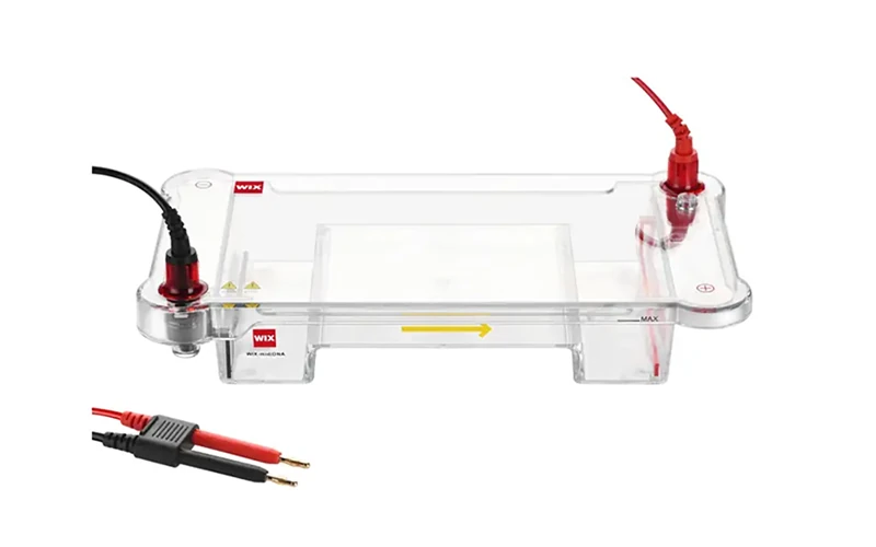
wix minidna horizontal electrophoresis cell for dna fragment analysis

How to Avoid Leaks, Gel Tray Deformation, and Electrode Failure in Horizontal Electrophoresis

If you regularly run agarose gels, you've probably encountered at least one of these problems: a leaking electrophoresis tank, a warped gel tray, broken platinum wires, or a failed run caused by loading samples in the wrong direction. While these issues may seem minor, they can waste valuable samples, delay experiments, and affect the reproducibility of DNA analysis. In busy teaching laboratories, research institutes, and diagnostic facilities, even small equipment-related problems can quickly add up to significant downtime.
View Details
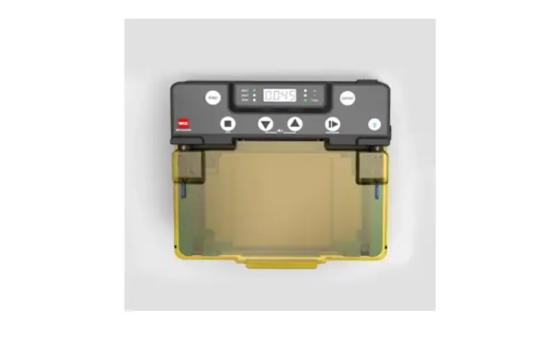
wix bluedna integrated horizontal blue light electrophoresis system with built in power supply and transilluminator.

Horizontal Blue Ray Electrophoresis System WIX-blueDNA: An Integrated Solution for Faster, Safer DNA Analysis

In the fast-paced world of life science research, time, accuracy, and sample integrity are everything. Whether you're running PCR product verifications, restriction digests, or RNA analyses, horizontal electrophoresis remains a cornerstone technique in molecular biology labs. However, traditional setups often involve multiple standalone devices—an electrophoresis tank, a separate power supply, a UV transilluminator, and a gel documentation system—leading to fragmented workflows and increased risks of sample damage or user error.
View Details
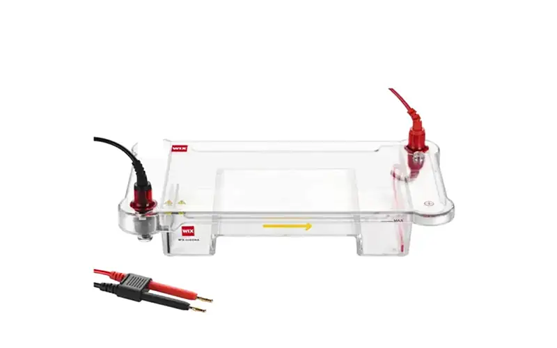
horizontal electrophoresis cell

Why Is the Horizontal Electrophoresis Cell WIX-midiDNA a Practical Choice for DNA and PCR Analysis?

In the field of life science, few techniques are as foundational as electrophoresis. Whether you are separating DNA fragments after PCR or analyzing RNA samples, a reliable horizontal Electrophoresis system is essential. Among the many options available, the Horizontal Electrophoresis Cell WIX-midiDNA stands out as a versatile and high-throughput tool designed for modern molecular biology laboratories. This article explores why this system is a practical choice for routine DNA Electrophoresis and PCR electrophoresis workflows.
View Details
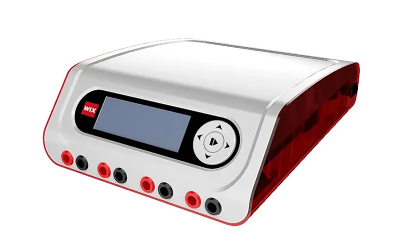
electrophoresis power supplies 1

Electrophoresis Power Supplies Explained: Real-World Applications, Tips & Expert Insights

In the realm of life sciences and molecular research, electrophoresis stands as a foundational technique, enabling the separation and analysis of charged molecules like DNA and proteins. While many labs focus on reagent quality or gel preparation, the power supply's ability to deliver stable electrical output is often the deciding factor between reproducible, publishable results and costly, time-consuming failures.
View Details

Choosing the Right Horizontal Electrophoresis Cell for Your Sample Volume

Sample throughput varies significantly between horizontal electrophoresis cells, and matching capacity to your daily workload is one of the first decisions to get right. Our guides compare compact options like the WIX-miniDNA, suited for smaller batches of up to 32 samples, against higher-capacity systems like the WIX-midiDNA, which supports up to 108 samples across four interchangeable gel tray sizes. These articles also walk through how comb thickness and well configuration affect sample loading, so you can match tray size and comb selection to routine checks versus larger PCR batches.

Preventing Leaks, Gel Tray Deformation, and Electrode Failure

Buffer leakage, warped gel trays, and broken platinum electrodes are some of the most common frustrations in day-to-day horizontal electrophoresis work. Our troubleshooting guide breaks down what causes these issues, from aging rubber gaskets to undersized platinum wire, and explains how design features like snap-fit electrode assemblies and heat-resistant gel trays address them directly. If your lab is dealing with recurring maintenance issues or inconsistent DNA band results, this is a good place to start.

Moving to an Integrated System for Faster DNA and RNA Workflows

Traditional horizontal electrophoresis setups often mean juggling a separate tank, power supply, and UV transilluminator, which adds handling time and exposes samples to UV light that can damage DNA. Our article on the WIX-blueDNA covers how an all-in-one system with a built-in power supply and blue light imaging simplifies this workflow, and where it fits for labs running PCR screening, cloning, or higher sample volumes across academic, pharmaceutical, or diagnostic settings.

Frequently Asked Questions (FAQ)

Q: What’s the difference between horizontal and vertical electrophoresis?

A: Horizontal electrophoresis cells are designed for nucleic acids—DNA and RNA—run on agarose gels, covering applications like PCR verification and restriction digest analysis. Protein separation techniques like SDS-PAGE use vertical electrophoresis systems instead, so the right choice depends on whether you’re working with nucleic acids or proteins.

Q: Why does my gel tray warp or deform when I pour hot agarose?

A: This usually happens with trays made from standard plastic, which can lose their shape at temperatures above 60–70°C, forcing you to wait for the agarose to cool before pouring. Heat-resistant tray materials can tolerate much higher temperatures, letting you pour molten agarose directly into the tray without deformation.

Q: Is blue light imaging actually safer than UV for DNA samples?

A: Yes. UV light, particularly at 254 nm, can cause DNA damage that reduces downstream cloning or sequencing efficiency. Blue light imaging (470–490 nm) excites the same fluorescent dyes without that damage risk, which matters if you plan to recover DNA fragments after the run.

Contact Form
Get A Quote
Contact Form