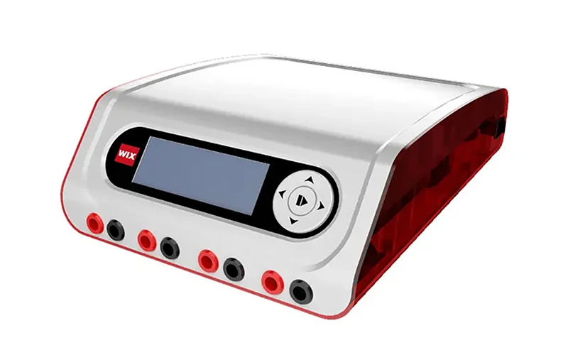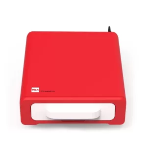
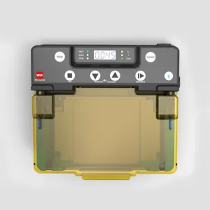
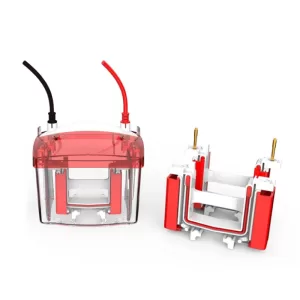
Do You Have Interest in Electrophoresis Tanks?
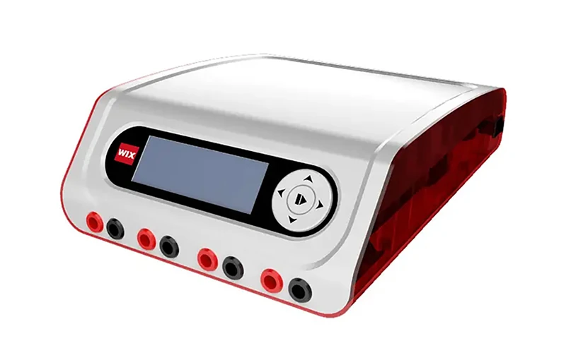
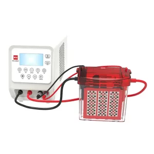
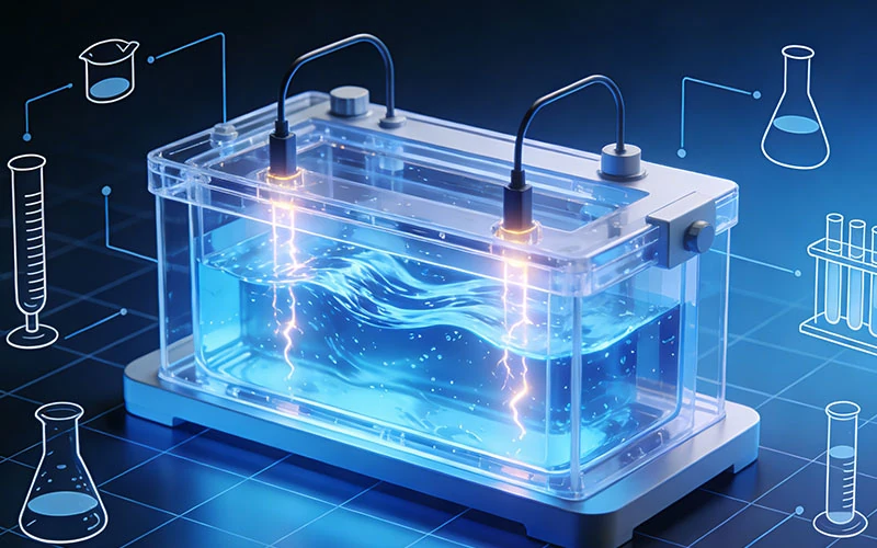
An electrophoresis tank is a core device used to hold electrophoresis buffer and samples in electrophoresis experiments. Its design and performance directly affect the accuracy and reproducibility

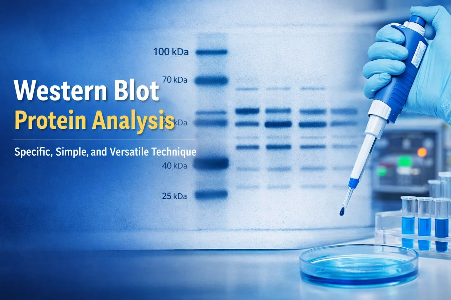
Conventional gel imaging systems are widely used for routine electrophoresis imaging, such as capturing DNA and RNA bands from agarose gels. However, different applications require different detection capabilities. Western blot chemiluminescence imaging, for example, involves weaker light signals and requires higher sensitivity and better background control than standard fluorescence imaging.
Multi-function gel imaging systems combine nucleic acid gel imaging, protein gel documentation, and chemiluminescence detection into one imaging platform, providing a more flexible solution for diverse molecular biology workflows.
A multi-function gel imaging system is an imaging platform used to capture, visualize, and analyze biological samples after electrophoresis. Unlike basic gel imaging equipment that is mainly designed for nucleic acid visualization, multi-function systems are capable of handling different types of biological signals, including DNA/RNA bands, protein gels, and chemiluminescent signals from Western blot experiments.
These systems combine multiple imaging modes within one platform, allowing researchers to perform gel image acquisition and analysis for a variety of molecular biology applications. By integrating fluorescence imaging, visible light imaging, and chemiluminescence detection technologies, multi-function gel imaging systems provide a more comprehensive approach for documenting experimental results in life science research.
Traditional gel imaging systems are commonly used for routine fluorescence imaging, particularly for visualizing nucleic acid bands after electrophoresis. These systems can meet many standard laboratory requirements, but advanced applications such as Western blot chemiluminescence detection may require higher sensitivity, longer exposure capability, and better control of background signals.
As imaging applications expand, multi-function gel imaging systems have evolved to support multiple detection methods within a single platform. By combining nucleic acid gel imaging, protein gel imaging, and chemiluminescence detection, these systems provide greater flexibility for laboratories handling different types of biological samples.
A multi-function gel imaging system works by converting biological signals from electrophoresis samples into digital images that researchers can visualize, analyze, and document. Depending on the sample type, the system uses different imaging methods to capture DNA, RNA, or protein signals.
The imaging process usually involves five main steps:
Before imaging, biological samples are separated through electrophoresis and prepared with appropriate detection methods.
For DNA and RNA analysis, agarose gels are commonly stained with fluorescent dyes to make nucleic acid bands visible. For protein analysis, PAGE gels may use colorimetric or fluorescent staining methods. In Western blot experiments, chemiluminescent substrates are applied to generate light signals from target proteins.
Different biological samples require different illumination methods.
For nucleic acid gels, UV or blue light excitation is commonly used to visualize fluorescent DNA bands. For protein gels, white-light illumination helps researchers observe stained bands. In chemiluminescence imaging, the system detects light signals generated directly from the Western blot membrane without external illumination.
After illumination or chemiluminescent signal generation, the imaging system collects the emitted light through a sensitive camera.
For applications such as Western blot imaging, signals can be very weak, so high-sensitivity imaging sensors and optimized optical systems help capture clearer images during longer exposure times.
The captured light signals are converted into digital data by the imaging sensor.
Advanced gel imaging systems use sensitive cameras to record differences in signal intensity, allowing researchers to distinguish between stronger and weaker bands within the sample.
After image acquisition, dedicated software processes the digital image for further analysis.
Researchers can measure band intensity, compare sample differences, estimate molecular characteristics, and prepare images for research records or scientific publications.
Accounting for the largest share of end users, these systems are essential tools across university departments—such as Life Sciences, Basic Medicine, and Agriculture—for studying gene expression, protein structures, and molecular mechanisms.
Within pharmaceutical enterprises and biotechnology service providers, they facilitate drug discovery, biomanufacturing quality control, and contract research by ensuring precise quantitative analysis of biomolecules.
These imagers are widely deployed across general hospital departments (including clinical laboratories and pathology) as well as third-party medical testing facilities for molecular diagnostics and biomarker evaluation.
Various niche sectors rely on gel imaging for critical compliance and testing tasks, ranging from agriculture and food safety verification to forensic identification, judicial investigations, and anti-doping analysis.
This device integrates nucleic acid gel imaging, protein staining analysis, and ECL Western blot imaging into a single unit, saving valuable lab space and reducing hardware maintenance costs.
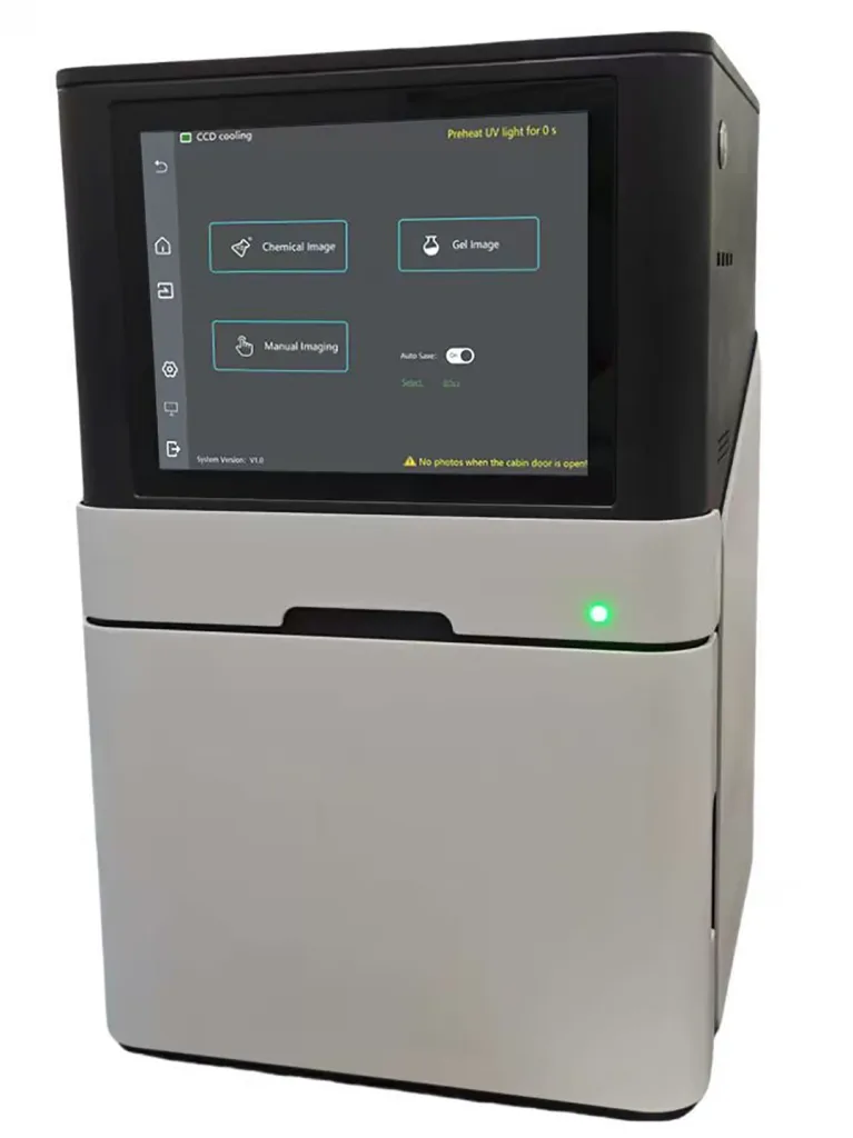
It can achieve detection limits in the low picogram (pg) to femtogram (fg) range combining an F0.95 lens, -35°C TEC cooling technology, and a peak quantum efficiency (QE) of 90%. This offers a clean, digital alternative to traditional X-ray film development, eliminating the need to handle chemical waste from darkrooms.
A double-layer light avoidance front door prevents room lighting from leaking into the darkroom. With low dark current (0.002e-/pixel/sec), background pixel gray levels can remain stable making it easier to distinguish closely spaced target bands.
The built-in computer and 12.1-inch capacitive touch screen simplify multi-user operations. Researchers can acquire images and run band densitometry analysis directly at the bench without queuing for external workstations.
Evaluate Camera Sensitivity
When selecting a gel imaging system, consider key camera specifications such as quantum efficiency, full well capacity, and A/D conversion depth. These factors influence the system’s ability to capture weak biological signals, especially in low-light applications such as chemiluminescence imaging.
Consider Cooling Performance
Chemiluminescence imaging often requires longer exposure times, making thermal noise control an important factor. Camera cooling technologies, such as TEC cooling, can reduce sensor dark current and help maintain image quality during weak signal detection.
Confirm Light Source and Filter Compatibility
Different experiments require different illumination methods. Check whether the system supports suitable light sources and filters for your applications, including UV, blue light, or white light imaging. Flexible optical configurations can help accommodate future experimental needs.
Modern multi-function systems incorporate Trans-UV (302/365 nm), Trans-White, Epi-Blue (470 nm), and darkroom chemiluminescence modes. This configuration lets researchers handle agarose gels, SDS-PAGE gels, and ECL Western blot membranes all inside a single benchtop darkroom.
Chemiluminescent signals emit a faint glow over an extended period. Without a cooling system, the heat-induced dark current generated would obscure low-intensity bands. Dual-stage TEC cooling technology lowers the sensor temperature to 35°C below ambient, reducing dark current to 0.002 e⁻/pixel/sec and ensuring clear signal contrast during long exposures.
Multi-function gel imaging systems have become an important imaging solution for laboratories that need to analyze different types of biological samples, from nucleic acids to proteins. By integrating multiple imaging capabilities, these systems help simplify experimental workflows and provide a more efficient way to capture, analyze, and document electrophoresis results.
From imaging principles and application requirements to key selection factors, understanding the technologies behind gel imaging systems can help researchers choose a suitable solution for their specific workflows. With continued development in camera sensitivity, optical design, and imaging automation, gel imaging platforms will continue to support research, biotechnology, clinical testing, and other biological applications.